
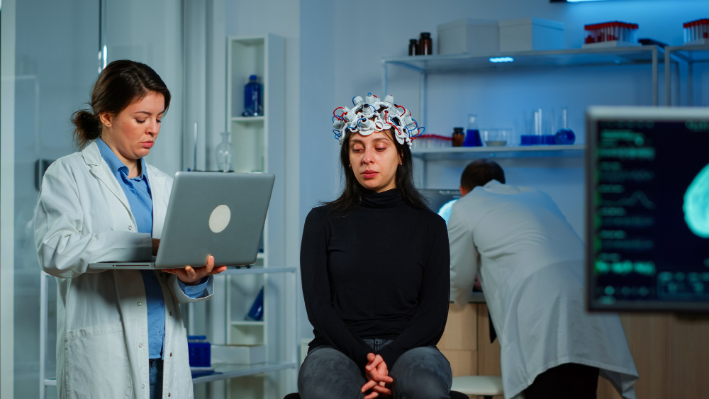
The evidence has been quietly building for years inside an MRI scanner at a research facility, between a set of pre-flight baseline scans and a post-mission medical evaluation. From a distance, the pictures don’t appear dramatic; they are precise, grayscale cross-sections of the human brain that are marked with dates and mission durations. However, it took some time for researchers to fully comprehend what they discovered when they started aligning those images skull-by-skull and comparing pre-flight and post-flight positions: the brain had moved. Not in a symbolic sense. Not in the sense of altered processing or cognitive change. physically relocated. moved upward and backward inside the skull, changing its shape in relation to the surrounding bone in ways that corresponded exactly with the astronaut’s duration in space.
Building on ten years of NASA-funded neuroimaging research, a University of Florida study released in March 2026 examined brain MRI scans from 26 astronauts who had been in space for a few weeks to more than a year. The team separated the brain into more than 100 distinct regions and monitored each one independently rather than treating the brain as a single entity and calculating average changes. Because specific movements on opposing sides of the brain cancel each other out when averaged, the method uncovered patterns that previous whole-brain analyses had entirely missed.
| Human Brain Changes in Space — Key Research Data | |
|---|---|
| Key 2026 Study | “Living in space can change where your brain sits in your skull” — Tianyi (Erik) Wang & Rachael Seidler |
| Lead Institution | University of Florida (Department of Applied Physiology & Kinesiology); related MIT spatial computing research — MIT Picower Institute |
| Published | March 7, 2026 — The Conversation / Space.com |
| Participants | 26 astronauts — missions ranging from a few weeks to over a year |
| Key Finding | Brain shifts upward and backward inside the skull after spaceflight; larger shifts in longer missions |
| Largest Regional Shift Observed | Over 2 millimeters upward in some areas near the top of the brain — in year-long mission astronauts |
| Recovery Timeline | Most shifts return to normal within 6 months post-return; backward shift recovers more slowly |
| Ventricular Volume Changes | Significant enlargement after long missions; rate over 3x faster than normal aging |
| Brain White Matter Changes | Reduced fractional anisotropy; structural changes in corticospinal tract, superior longitudinal fasciculus, cerebellar peduncles |
| Visual System Risk | Spaceflight-Associated Neuro-ocular Syndrome (SANS) — flattening of the eye, optic disk edema, vision changes |
| NASA Relevance | NASA Human Research Program — studying space health impacts for Artemis and future Mars missions |
| Key Comparison Condition on Earth | Idiopathic intracranial hypertension (IIH) — similar fluid pressure symptoms |
| Systematic Review Coverage | 20 neuroimaging studies reviewed; all using MRI — structural, diffusion-weighted, and functional |
| Prior Groundbreaking Study | MUSC/NASA study (2017) — confirmed upward brain shift and central sulcus narrowing in 94% of long-duration astronauts |
In astronauts who had finished a year-long mission, some regions close to the crown of the brain had moved upward by more than two millimeters. Two millimeters doesn’t seem like much. It is not inside the highly compressed area of a human skull. Movement and sensation-related areas showed the biggest displacement; these are precisely the systems astronauts need to operate correctly when they return to gravity.
Although the implications of this are complex, the underlying physics is simple. The body’s fluids are constantly drawn downward by gravity on Earth. Under that continuous directional pull, the brain, floating in cerebrospinal fluid inside the skull, achieves a stable equilibrium. That equilibrium vanishes when gravity is removed. Body fluids move toward the head, both internally as a redistribution of cerebrospinal fluid that the brain lacks an evolved mechanism to anticipate or fully compensate for, and externally as the puffy faces astronauts experience during the first few days of a mission.
The brain is more buoyant. Compression occurs in the fluid spaces at the top of the skull. Structures that were balanced against gravity’s pull abruptly encounter a different set of forces, which causes them to deform. The changes are less significant and recover faster for brief missions. The changes are greater, more enduring, and sometimes still detectable six months after the astronaut returns to Earth for missions lasting six months or longer, which is currently the typical length of an International Space Station rotation.
Although this science is not wholly novel, the accuracy of recent discoveries differs significantly from what was previously available to researchers. According to a 2017 study from the Medical University of South Carolina, 94% of long-term astronauts had narrowing of the central sulcus, which is the cortex’s groove that divides the parietal and frontal lobes. MRI cine clips also clearly showed an upward brain shift. In addition to the discovery that opposing movements in different hemispheres had been masking one another in previous whole-brain analyses, the 2026 study adds a detailed regional map of precisely which parts of the brain are moving and by how much. The science was concealed within its own methodology.
Twenty neuroimaging studies on astronauts using structural MRI, diffusion-weighted imaging, and functional MRI were examined in a systematic review that was published in Brain Imaging and Behavior. All of those studies paint a complex picture. During long-duration missions, ventricular volume increases significantly at a rate more than three times faster than normal aging.
Crucially, the enlargement does not rapidly reverse upon return. The superior longitudinal fasciculus, the corticospinal tract, and the cerebellar peduncles—all pathways involved in balance, motor coordination, and sensory integration—all exhibit white matter alterations. The brain’s connectivity patterns also change after flight; some networks exhibit decreased connectivity, while others exhibit increases that scientists believe are compensatory reactions, the brain rewiring itself to continue functioning under odd circumstances. After landing, some of those changes are reversed. Some last for months, and it’s still unknown which ones last forever.
Perhaps the most clinically concerning effects are those on the visual system. NASA discovered that a large percentage of long-duration astronauts suffer from a condition known as Spaceflight-Associated Neuro-ocular Syndrome, or SANS. Increased pressure from the upward fluid shift causes the optic disc to swell, the back of the eye to flatten, the light-sensitive retinal layer to fold, and the visual system to become farsighted. After returning to Earth, these changes have continued for some astronauts. Although the precise mechanism linking intracranial fluid dynamics to vision loss is still unclear, NASA has designated it as a top research priority due to the strong correlation.
It’s difficult to ignore the fact that ambition has influenced this research’s course just as much as curiosity. After a historic mission beyond the Moon, Artemis 2 brought its crew back to Earth in April 2026. The next stated destination is Mars; the trip will take at least six months each way, and there is no way to return quickly.
After six to twelve months in low Earth orbit, under the relative protection of Earth’s magnetic field, the brain changes observed in ISS astronauts may be less than those that would happen during an interplanetary transit where they would be exposed to greater radiation and more isolation. No one has yet provided a definitive answer to the question of whether existing countermeasures, such as drug interventions, exercise regimens, and fluid management techniques, are adequate to safeguard the brain during a multi-year mission. The readable version of a problem whose complete dimensions are still being mapped is the two millimeters of shift that can be seen on an MRI.